Overview
This proposal aims to improve Medical X-Ray Imaging using Machine Learning (ML) and Sparse Representation Theory(SRT). The project’s main goal is to combine, state of the art image enhancing techniques in x-ray medical imaging focusing in mammography, and feed them to specially designed ML algorithms leading to novel Computer Assisted Diagnosis (CAD) tools. The emerging state of the art research field of ML, actually provides computers the ability to “learn” without being explicitly programmed. ML has been greatly evolved the last decade due to the immense computational capabilities of the new generation of Graphics Processing Units’ (GPU). First efforts to apply ML in medical x-ray imaging have taken place, with promising results, as in the case of chest x-ray imaging (Zhiyong Lu et al., 2017), computer aided detection of tuberculosis (Philipsen et al., 2015), detection tumour growth within the human skeleton (Idota et al., 2016) and very recently a first attempt for the case of mammography was done by the Department of Physics of Complex Systems in Hungary (Ribli et al., 2018) using the INbreast (Moreira et al., 2012) full-field digital mammographic dataset. The application of ML techniques in medical imaging is therefore appearing as a very promising, novel, state of the art research field for the years to come. On the other hand, SRT founded within the last decade, revolutionized the signal and image processing approaches. The application of SRT-based methods into “classical” problems that puzzles scientists for decades, such as signal or image denoising, provided surprisingly good results. Furthermore, SRT-based methods known as Compressive Sampling (CS) have challenged even the well-established sampling theorem of Shannon. The combination of these two fields is expected to provide new effective tools for image enhancement and diagnostic capabilities in medical imaging.
The application of both ML and SRT in medical imaging is still at an early stage, designating it as an open and very promising field of research and innovation. Considering, in particular, the X-Ray medical imaging field, it is expected that advanced, SRT-based techniques for image processing and enhancement along with ML based analysis and classification, will lead to superior image quality, with improved diagnostic potential.
The proposed research will focus on adaptation, evaluation and testing of a series of medical image enhancement and processing techniques, as well as advanced image analysis approaches. A software library is to be developed, with algorithms for de-noising (filtering), edge and contrast enhancement, super resolution and ML based CAD. This is an innovative approach, where medical images will be enhanced using state of the art SRT techniques and then will be fed to ML algorithms in order to automatically detect lesions and malignancies in Breast imaging. Expect from the innovating approach of combining of SRT and ML, the use of ML for the detection of malignancies in breast x-ray imaging, is a very promising, not yet thoroughly investigated, research field. For the purpose of this research, the existing in-house Monte Carlo XrayImagingSimulator (Bliznakova, 2003; Bliznakova et al., 2010; Lazos et al., 2003, 2000).developed by Biomedical Technology Unit (BITU) and used in numerous research schemes and projects from teams all around the world, will be used. Applications of this simulator, which has been developed the last 15 years, are not limited in the field of medical imaging, as in the case of project QUICOM, where it was employed for the non-destructive testing of airplane carbon fibre parts.
The specific objectives of the proposed project are:
- Development/Implementation of an ML based CAD algorithm for the automatic detection of lesions and malignancies in Breast imaging
- Development of an X-ray Medical Image Enhancement Library that will comprise the following algorithms (utilizing the SRT) for:
- Image denoising (filtering) in 2D and 3D imaging
- Super resolution for improving the resolution of medical images
- Image contrast and edge enhancement
- Design and development of software phantoms for the production of synthetic data using X-Ray Imaging Simulator and physical phantoms using conventional and 3D printing techniques.
- Validation of the developed algorithms using real data.
- Conduction of an evaluation study for the performance evaluation of the derived ML based CAD algorithms versus the diagnosis provided by medical experts
In order to achieve these goals, the duration of the project is set to 36 months and a multidisciplinary research team of highly experienced researchers, has been assembled. The members of the team are affiliated with research groups that are highly experienced in these fields and have long track record in the areas covered by the proposed project.
A successful outcome of the proposed project will lead to better tools for breast lesion and malignancies diagnosis, creation of powerful SRT and ML tools for future research and will allow involved groups and investigators to expand the application of this approach to other fields of X-ray Medical Imaging and to remain in the front end of these research fields.
Implementation
The overall structure of the project’s work plan consists of 5 Work Packages (WP) shown in Figure 1. In particular:
1. WP1: extensive literature review and determination of the state of the art concerning the available technologies, overall design of the system. Outcome: report on the state of the art and project’s system plan.
2. WP2 (Simulation Phase): At the middle of WP1, ML and SRT algorithms development phase along with simulations using XrayImagingSimulator, will start to ensure that all needed data will be available for the development of the algorithms. A milestone at the middle of WP2 when a first version of the algorithms will be ready and tested based only on simulation data. At this point, the CUDA (GPU) version of the developed algorithms will also start. Outcomes: software phantoms, (some designed to be also physically built), the developed of refined and validated via simulation algorithms, based on initial application results from (WP3).
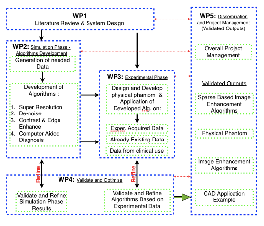
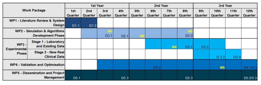
3. WP3 (Experimental Phase): begins in the middle of (WP2) where, a first version of all the proposed algorithms will be available along with the software phantoms. In Stage 1 of WP3 actual physical phantoms will be created using conventional and 3D printing techniques and will be used for x-ray imaging. The algorithms will then be applied on the produced experimental data and, based on the outputs, the algorithms will be refined. The CAD’s system training will be refined using all the labelled experimental data produced. Algorithms will be also applied in existing data from previous collaborations and experiments of the BITU team and PI in Elettra synchrotron facilities and from collaborating hospitals. In the 2nd stage of WP3 new real clinical data will be acquired. All image enhancement algorithms will be tested and validated using these data, and all necessary improvements will be done. The ML CAD system, will now enter its final training phase, where after the successive augmented training starting with simulated and then experimental data, will be now fed and trained using labelled real clinical data from available open access mammographic datasets (Moreira et al., 2012) (Suckling et al., 2015) along with data available in the BITU team and collaborating hospitals. An evaluation study will take place where images will be given to experts to analyse them and comparison with the ML CAD outputs. Outputs: physical phantoms, experimental data and algorithms tested, refined and retrained with real data.
4. WP4 (Validation and Optimization): Follows the beginning of WP2, a continuous Validation and Optimization procedure will run in parallel with WP2 and WP3 until the end of the project. All the produced algorithms and data will be validated and, when appropriate, redesigned and refined. This approach has been chosen in order to make sure that any problem that may occur, will be detected and all needed actions will be done at an early stage. Running of WP4 in parallel from the beginning of the project, will also allow the direct comparison of the results acquired by the simulation and experimental phases and switching from simulation to experiments and vice versa in order to validate and refine the produced algorithms. This continuous monitoring and evaluation will ensure the positive outcome of the project. Output: final versions of the derived algorithms.
5. WP5 (Dissemination & Management): dissemination actions such as paper submission in international peer reviewed journals, conferences and presentation of work, along with a web site of the Project. A Workshop on project’s outputs and related scientific fields will take place in the facilities of Patras University, with invited researchers from Greece and EU. Project management will take place in WP5 and will be running s throughout the whole duration of the project.
Research Team
Biomedical Technology Unit (BITU, bitu.upatras.gr), is headed by Emeritus Prof. N. Pallikarakis, is recognized as one of the pioneers and major contributors to the development of Digital Tomosynthesis (DTS). The research has been directed towards the development, validation and implementation of a DTS imaging prototype, based on the Multiple Projection Algorithm (MPA), developed in the early 80’s and patented in 1994. It was then applied into a Cone Beam Computed Tomography (CBCT) formulation. Concurrently, research was oriented towards noise removal algorithms, motion correction methods and simulation. In the end of 90s, the basic Radiographic Simulator was developed along with a realistic Breast Model, resulting in a Mammographic Simulator and the Dual-Energy Simulator. The Radiographic Simulator has evolved to a dedicated X-ray Imaging Simulator that incorporates Monte Carlo methods for simulation of irradiation transport. Further on a Monte Carlo based simulator of the Radiotherapy Treatment chain was developed and validated. BITU’s team possess a strong experience in modelling and simulation with in house developed software and hardware tools. The team has performed simulations in testing new technologies of treatment of neck and head cancers, imaging of the breast cancers, brain cancer imaging simulation and dual energy techniques. All this experience will be used in order to manage and guide the proposed research project into combining the state of the art computational and signal processing techniques with medical imaging. BITU will provide all the support needed in infrastructure, project management experience, available personnel and technological equipment along with available mammographic data that have been acquired either experimentally from BITU’s experiments in the Elettra synchrotron facility or real clinical data from collaborating hospitals and physicians, in order to achieve project’s stated objectives. This research activity lead to more than 100 Publications in peer reviewed journals and 12 PhD thesis on this area over the last 25 years.
Emeritus Prof. N. Pallikarakis, former Director of the BME postgraduate program, Author of more than 130 scientific papers and Project Coordinator of more than 30 national and European R&D projects, National delegate for the MD Standing Committee (DG III), the HCF of CEN and elected member of the International Academy of MBE. He will be the overall coordinator of the project and will participate in all WP (without getting paid for his contribution)
Dr. Aristeidis Dermitzakis, holds a PhD in Biomedical Engineering in the field of x-ray imaging, will be responsible for the scientific and technical coordination of the overall project, and more specifically for the ML, simulation and evaluation tasks of the project.
Dr. Natassa Daskalaki, holds a PhD in Biomedical Engineering in the field of Breast Imaging, will be responsible for the breast imaging simulation tasks and experimental data image characterization for the algorithm training tasks.
Dr. Dimitris Ampeliotis, holds a PhD in Signal Processing, will be responsible for the SRT related tasks of the project.
Dr. Anthi Malliori, holds a PhD in Biomedical Engineering in the field of Tomosynthesis for Breast Imaging, will be working on breast imaging simulation tasks and experimental data image characterization needed for algorithm training tasks.
Dr. Evangelos Vlachos, holds a PhD in Signal Processing and is currently a post doc researcher in University of Edinburgh School of Engineering, which will be a collaborating institution, offering great advantage to this project’s overall expertise. He will be working for the SRT and ML related tasks of the project.
1η Προκήρυξη ερευνητικών έργων ΕΛ.ΙΔ.Ε.Κ. για την ενίσχυση των μελών ΔΕΠ και Ερευνητών/τριών και την προμήθεια ερευνητικού εξοπλισμού μεγάλης αξίας

Αθήνα, 07.12.2018 | Α.Π. 7518
Πρότυπα Έγγραφα – Προτάσεις Κατηγορίας Ι ή ΙΙ (Β΄ Φάση Υποβολής)